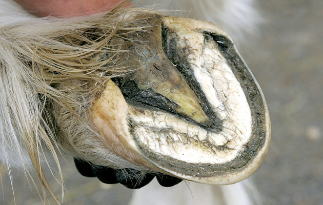
Much of what is known about the equine foot has its historical origins in the work of farriers and blacksmiths. Early veterinary texts discuss a number of conditions familiar to us today, such as navicular disease (grogginess), laminitis (fever in the foot), canker and corns.
Advances in veterinary science have deepened our understanding of the internal structures of the foot, revolutionising the way farriers approach hoof care. We now know that a good working relationship between vet and farrier is essential for soundness in the horse.
But what can the modern veterinary world teach farriers?
With the discovery of X-rays in the late 19th century, it wasn’t long before the X-ray tube was pointed at the horse’s foot. X-rays (radiographs) primarily diagnose bone-related conditions. A change of around 30% bone mineral density may be required, however, before signs are visible on a radiograph image.
Not only does this mean that early disease changes may not be detectable by the naked eye on radiographs, but also that clinical signs — namely lameness — do not always correlate with what the images show.
As well as assisting with a diagnosis, radiographs are useful for the farrier in the assessment of foot balance. Through the use of good medio-lateral (from the side) and horizontal dorso-palmar (front to back) radiographs, the farrier can appreciate how external “landmarks” in the hoof construction are related to internal structures.
Although radiography can inform on some conditions involving soft tissues of the foot, it is the availability of magnetic resonance imaging (MRI) that has completely opened up our ability to see inside the hoof.
MRI — the inside story
MRI should never take the place of a good clinical assessment and veterinary diagnostic work-up. Static and dynamic examination of the whole horse should be performed by vet and farrier (ideally both together). Evaluation of the horse’s conformation, as well as palpation and visual assessment, should be undertaken and related to the presenting signs.
The farrier will then check the feet and limbs for obvious problems or damage, and assess foot balance and shoeing in relation to movement. Assessment is performed at both walk and trot, with a lameness value often being assigned by the vet.
Seeing the horse walk is particularly important for the farrier, because how the foot lands — flat or laterally, for example, or toe or heel first — is related to foot balance. Heavy landing on one side or the other can eventually lead to foot-related lameness.
From the vet’s perspective, localisation and response to diagnostic analgesia (nerve blocks) should always be performed to confirm the source of lameness as within the foot.
Horses with forelimb lameness originating in the foot may look “shoulder lame”, as they generally pull up and away at the shoulder/neck region. It is important to be focusing on the right area from the start of the treatment.
Before rushing the horse into the MRI scanner, good-quality foot radiographs are essential. A number of conditions can be diagnosed without MRI and dealing with obvious foot imbalance may be necessary before progressing to advanced imaging techniques such as MRI.
That said, even in cases where a diagnosis has been reached by radiography (such as navicular disease), MRI can provide information that radiography simply cannot — not only about the soft tissues, such as tendons and ligaments, but also about what is going on inside the bones.
This additional information can inform vet, farrier and owner on treatment options as well as likely prognosis. Indeed, with conditions of the navicular apparatus, for example, radiographs often severely underplay the extent of changes present.
Collateral damage
One area where MRI has been instrumental is the recognition of the importance of injuries of the collateral ligaments of the distal interphalangeal (coffin) joint.
These injuries are often diagnosed in sport horses, possibly related to the demands of their competition workload on various surfaces. There may also be an association of foot balance and conformation, where medio-lateral imbalance and rotational abnormalities may lead to stress across the joint and hence the ligaments.
Collateral ligament injuries can present as an acute lameness, sometimes seen if a horse suddenly stops and twists at a fence or gate, for example. They commonly present as a chronic, insidious lameness, however, often made worse when the horse is worked — particularly when lunged on a circle.
Diagnosis may be made through ultrasound examination, although full evaluation of the ligament can only be obtained via MRI.
A treatment plan is then made in conjunction with vet, farrier and physiotherapist.
Agreement on best management varies and may include a period of rest and controlled exercise (usually over three to six months), pain relief (systemically or via medicating the coffin joint directly) and physiotherapy treatment (through the ligament at the coronary band or via the hoof wall).
It is agreed, however, that corrective farriery is essential to address any underlying foot imbalance and reduce abnormal stresses across the joint and through the collateral ligaments during the recovery period.
The take-home message here is that if your horse is diagnosed with a soft tissue injury to the foot, make sure that the farrier is involved as early as possible in the management of the case.
Ref: Horse & Hound; 25 June 2015
