A specialised equine CT scanner — the first in the UK — has been unveiled at the Royal Veterinary College (RVC).
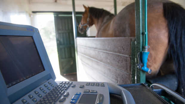
The new computer tomography (CT) scanner, which is at the RVC’s equine hospital in Hertfordshire, is 10cm wider than the standard size (75cm). This means that most of a horse’s neck fits within it.
Although the hospital has been performing CT scans for years, this new scanner will allow vets to easily scan more of the horse than they have previously been able to.
This will help with diagnosis, improve understanding of neck problems, and enable development of new treatments.
“We are very excited to introduce this innovation in equine imaging,” said Professor Renate Weller of the RVC.
“This is a great opportunity to improve our diagnostic ability for neck problems in the horse and we expect that our understanding of neck problems will improve tremendously.”
The RVC was the first equine hospital to install a CT scanner for horses in 2003. In 2009, the college changed its CT set-up so horses could be examined while they were standing.
This removed the need for general anaesthetic in order for a horse’s head and most of his neck to be examined.
Using a specially adapted platform, the head and neck can be easily moved in and out of the scanner.
“The RVC recognises that there is a risk with general anaesthetic and is keen to avoid administering it unnecessarily,” said a spokesman.
He added the new scanner builds on the “major step” of being able to examine horses while standing.
More than 80% of cases undergo at least one imaging procedure at the equine hospital, such as X-rays, CT scans or magnetic resonance imaging (MRI).
Related articles:
- Sticking stifles: what you need to know *H&H VIP*
- How do we know what the best veterinary treatment for a horse is? *H&H VIP*
- It shouldn’t happen to a vet (but it does…)
CT and MRI scans give virtual images that can be used to create a 3D picture of part of a horse.
This makes it possible to see very small, but clinically significant injuries and abnormalities.


