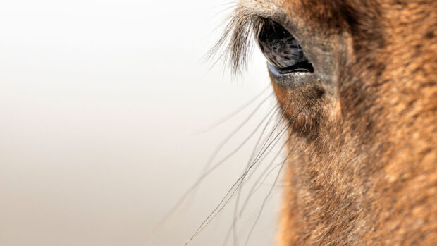
Ria Chalder MRCVS looks at some of the most common eye problems found in horses.
1. Eyelid laceration
Eyelid lacerations occur relatively frequently in horses when they catch their eyelid on a sharp object in the stable after rapidly lifting their head in response to a noise or perceived threat. Your vet will usually need to sedate the horse in order to determine the extent of the damage to the eyelid and the best way to repair it. Many horses that lacerate their eyelid will also have damaged the surface of their eye in the process (a corneal ulcer), so your vet may stain the surface of the eye with fluorescein dye, too.
How is it treated?
Eyelid lacerations should be regarded as an emergency and your vet called as soon as possible, so repair can be attempted before the damaged tissue begins to dry out and lose its blood supply. Many can be repaired with sutures (stitches) in the standing, sedated horse, but referral to a hospital for repair under general anesthesia may be required if there is extensive damage.
2. Eye problems in horses: immune-mediated keratitis (IMMK)
IMMK it is thought to be an immune-mediated disease whereby the horse’s own immune system attacks the surface of the eye. IMMK is not usually very painful, so signs can be subtle. Often a slightly cloudy white patch on the surface of the eye (referred to by vets as a “corneal opacity”) may be the only thing noticed at first.
A diagnosis of IMMK is made by ruling out alternative causes. Your vet may want to take some samples from the cloudy patch on the surface of the eye to rule out an infectious cause, such as bacteria or fungi, and to see what types of cells are present. Following this, a treatment trial of an immunosuppressive eye drop or ointment may be recommended in order to see if the eye responds to the treatment. Once a diagnosis of IMMK has been made, the disease can be further categorised into four types, based on how deep the disease is in the cornea. The four types are (in order of increasing depth): epithelial, superficial stromal, mid-stromal and endothelial IMMK. A piece of equipment called a slit lamp is usually required to determine the depth of the disease.
How is it treated?
Treatment will usually differ depending on the depth of the disease. Initial treatment usually consists of eye drops that suppress the immune response. This may be in the form of corticosteroids (steroids) or cyclosporine. Ruling out infectious causes prior to treatment is important as steroids should not be used if an infection is present.
In deeper stages of the disease, steroid injections in the tissue surrounding the eye, in addition to drops or ointment, may be used together. If the condition does not respond to topical treatment or a resistance to treatment develops over time, your vet may advise a surgical procedure called a superficial keratectomy, whereby the abnormal tissue is removed from the surface of the eye.
More recently, slow-release cyclosporine implants similar to those used in horses with ERU have been used in some cases of IMMK to remove the need for daily application of topical treatment. If left untreated, repeated episodes of IMMK can lead to the development of a scar on the surface of the eye.
3. Equine recurrent uveitis (ERU)
Equine recurrent uveitis (ERU) is a devastating autoimmune syndrome which causes inflammation inside the eye (uveitis), eventually causing blindness in approximately half of all horses affected. Research suggests that there may be a genetic component to the disease as it is seen more commonly in certain breeds such as Appaloosas and warmbloods. Environmental factors, injury and certain infections may also contribute.
ERU is diagnosed when there is inflammation seen within the eye, usually combined with pain and a history of similar episodes. There are three different types of ERU — the most common, “classic” ERU, is characterised by recurrent episodes of severe eye pain (squinting, tearing) and inflammation inside the eye lasting two to three weeks. “Insidious” ERU is typically less painful than the classic form, but low-grade inflammation still damages the internal structures of the eye over time.
Due to the more subtle signs, a diagnosis may only be reached quite late in the disease. “Posterior” ERU is least common. The inflammation and damage is in the back (posterior) of the eye, so can go unnoticed for some time.
How is it treated?
There is no cure, and treatment is aimed at reducing the frequency and severity of flare-ups. It usually involves anti-inflammatory medication in the form of immunosuppressive eye drops and oral anti-inflammatory medication, such as flunixin or phenylbutazone (bute). Atropine drops may also be used to help treat the pain and to open (dilate) the pupil. It is crucial that treatment is continued for several weeks, because the inflammation in the eye remains for a long time, even once clinical signs have resolved.
Stopping medication too early will likely result in another flare-up in the near future. If medical treatment is unsuccessful, there are a number of alternative options. For example, a small silicone device that slowly releases an immunosuppressive drug called cyclosporine can be implanted into the space surrounding the eye. The implants release the drug for up to five years.
Minimising potential triggers is important when managing horses with ERU — for example, decreasing UV and wind exposure. However, even with aggressive treatment, affected horses can go blind in a matter of years. Early involvement of a specialist veterinary ophthalmologist maximises the chances of managing the disease and prolonging the horse’s sight.
4. Eye problems in horses: corneal ulcers
Corneal ulcers occur when the surface of the eye (the cornea) is damaged. The prominent position of the horse’s eyes makes them more prone to injury, so corneal ulcers are a relatively common occurrence. They can be very painful, and horses with a corneal ulcer will often be found with their eye streaming and clamped shut. If your vet suspects a corneal ulcer, they will apply an orange dye called fluorescein to the surface of the eye. The stain will stick to damaged cornea only, highlighting the location and extent of any ulcer present.
How are they treated?
Most ulcers are simple scratches that heal quickly with appropriate treatment, which usually includes antibiotic drops to prevent infection while the ulcer heals. However, if an ulcer becomes infected with bacteria or fungi, it can take longer to heal. Inappropriate use of antibiotics over the past few decades has led to the emergence of multi drug-resistant strains of bacteria. For this reason, your vet may want to take a swab of the ulcer to determine which bug is growing and which antibiotic drop will be most effective against it. A medication called atropine may also be used which can relieve painful muscle spasm.
Some ulcers are slow to heal and may need the unhealthy tissue removing (debriding) with a cotton bud or a piece of equipment called a diamond burr. This is a relatively simple procedure, usually performed under sedation and with topical anaesthetic to numb the surface of the eye. Deep ulcers are considered an emergency and may require your horse to receive intensive around-the-clock medical treatment at a hospital, or even surgery to repair the damage.
- For unlimited access to our extensive online library of expert veterinary advice covering common equine aliments and conditions, subscribe to the Horse & Hound website
Found this useful? You might also enjoy reading these:

Equine eyesight questions answered, plus vets share advice on common vision problems to look out for
How to train, ride and manage a partially sighted horse: advice from owners with experience

All you need to know about the equine eye *H&H VIP*
Our new occasional series zooms in on the equine eye. Looking into this complex device can offer vital clues about

Equine recurrent uveitis (moon blindness) in horses
Discover the ideal fly mask to keep your horse comfortable all summer long

Subscribe to Horse & Hound magazine today – and enjoy unlimited website access all year round


