Few human adults have escaped without at least a filling or two as a consequence of tooth decay. While this concept is not something many of us would associate with our horse’s teeth, there is potential for decay in the equine mouth and the possibility that a filling technique may be needed to repair the damage.
Although they are made up of the same components, the structure of humans’ teeth is quite different to that of horses’ — not surprisingly, given our very different diets. In the human mouth, a layer of enamel completely covers the sensitive tissue beneath it, known as dentine, along with the pulp in the tooth core. Any defect in the enamel is likely to cause dental pain.
In the equine tooth, three types of tissue — enamel, dentine and a substance called cementum — are present on the occlusal (chewing) surface. If these hard, calcified tissues become demineralised, the organic component within can be destroyed — a process known as dental caries.
For a caries lesion (an area of demineralisation) to develop, there must be an interaction between a “substrate”, which is a carbohydrate in food, and the bacteria found in the sticky plaque that can build up on the tooth’s surface. The metabolism of carbohydrates by plaque bacteria primarily produces acids, which begins the demineralisation and caries lesion formation.
Because caries generally affects the cheek teeth, it is impossible to spot these lesions without a complete dental examination. Often a horse will show no clinical signs in the early stages, although halitosis, or smelly breath, always warrants further investigation, as does quidding, where the horse drops balls of semi-chewed food.

Peripheral caries
Breaking point
Caries can affect all the component equine dental structures, the cementum, enamel and dentine, and can be divided into two main categories: infundibular and peripheral.
Infundibular caries relates to the two infundibula — the cup-shaped enamel hollows present in the upper cheek teeth. The function of these is unknown, but the presence of enamel in the centre of the tooth does increase its grinding surface.
The cups are filled with cementum, which in the developing tooth spreads from its grinding surface towards the roots. In some cases, this process is incomplete, a condition known as cemental hypoplasia.
Caries first affects the cementum of the infundibula, when the condition is classed as grade one. It next involves the enamel (grade two) and then the dentine (grade three). Where the two diseased infundibula merge, a condition classed as grade four, this creates a central weakness in the tooth that is then vulnerable to a midline facture. Once the tooth has reached the point of fracture, extraction is the only treatment option available.
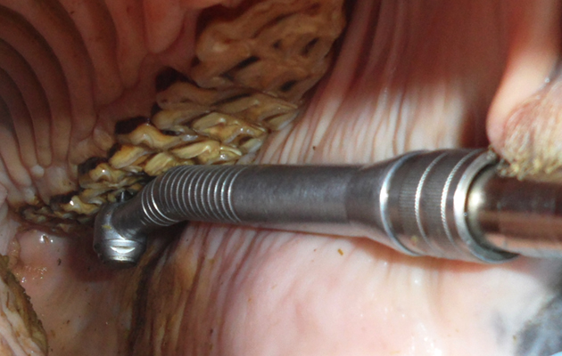
Identification of significant caries lesions requires a detailed examination under sedation, potentially using a viewing instrument called an oral endoscope for lesions that are very subtle. Some lesions are obvious, where food is packed in the infundibulum, while others may only be recognisable because tiny gas bubbles are leaking from the tooth. In suspected cases, further diagnostics, such as radiography (X-rays), may be performed to determine whether treatment by restoration is appropriate.
Restoration — or filling — involves debridement (cleaning) of any diseased material and food that is packed in the infundibulum. This is performed with a combination of high-speed dental drills; sharp-edged instruments called Hedstrom files and high-pressure water lavage (flushing), until the cavity is completely cleared.
Younger horses have longer teeth and therefore longer infundibula.
The cavities are then disinfected with mild bleach and their walls are roughened, or “etched”, with acid. These surfaces are then heat-sealed, a process known as bonding, before the infundibulum is layer-filled with a resin composite material. If etched, bonded and filled to the bottom of the infundibulum, these fillings should last the lifetime of the tooth.
Not all infundibular caries cases are suitable candidates for fillings, however. Any evidence of pulp disease must be investigated, as the tooth may require extraction due to disease around its roots.
Peripheral caries can affect all dental tissues on the non-chewing part of the tooth and is most commonly found in those at the rear of the lower jaw. In its mildest form, it involves only the cementum, progressing to the enamel and finally affecting the dentine before the tooth loses its structural integrity.
As the peripheral, or outer, cementum erodes, the teeth lose their rectangular shape. This can lead to food pocketing between them, which in turn exacerbates the problem as there is a greater amount of substrate present next to the caries lesions. It can also cause periodontal disease, an infection of the surrounding structures, which can be extremely painful.
The mainstay of treatment of peripheral caries is changing the horse’s diet to reduce the acid environment in the mouth, perhaps by swapping haylage for hay. Any sharp surfaces created on the teeth should be removed so as not to cause trauma. In some cases, where disease is advanced and has caused fracture, tooth extraction may be necessary.
Peripheral caries affects only the portion of tooth above the gum line. Once the cause has been removed, the continued eruption of the teeth may mean that they return to normal.
Is sugar to blame?
Atypical caries is similar in nature to peripheral caries, but less common and limited to the grinding surface of the tooth. Lesions affect the upper teeth, either side of the mid-cheek interdental space, and are often quite shallow.
Treatment of these cases is difficult and involves removing high-sugar, low pH foods from the horse’s diet.
The mouth can be flushed with a hose multiple times every day to stop food sticking to the teeth.
If an affected tooth otherwise appears normal, some specialists will debride it back to the healthy tissue, then bond resin composite to its surface to prevent progressive destruction of the tooth by decay. More severe and extensive cases may require multiple tooth extraction.
Caries can affect all ages and breeds. Although high amounts of sugar are thought to be a risk factor, this does not always follow.
The only practical management measure is to arrange routine oral examinations so that lesions can be spotted at an early stage. Because of the typically tucked-away location of caries lesions, however, a really detailed examination can only be performed under sedation.
Case study
A 12-year-old gelding referred to the dentistry clinic had bad breath and a history of quidding. An examination of his mouth revealed a midline fracture of tooth 209, which is located in the upper mid-cheek region. Food was packing deep into the centre of the tooth and fracture fragments were displaced to the inside and outside — with the outside fragment rubbing on the horse’s inner cheek and causing a large ulcer.
Following X-rays to rule out disease of the overlying sinuses, the tooth was removed via oral extraction. The extent of the fracture through both infundibula can be clearly seen.
Examination of tooth 109, on the opposite jaw, showed a deep infundibular caries lesion. This was classed grade two, affecting the cementum and enamel of one infundibulum — an earlier stage of the problem that affected the other tooth before it fractured. Luckily, this caries lesion could be debrided and filled. Three years later, the tooth remains healthy.
Ref Horse & Hound; 11 October 2018

